And now for something different… by Nienke Pannekoek

By Nienke Pannekoek (Leiden University)

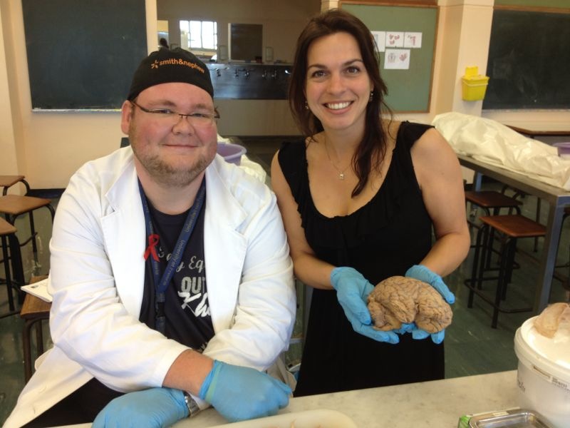
Having studied the brain for several years using MRI and by pictures in study books, getting the opportunity to take part in a brain dissection was something I had been hoping to experience for a long time.

Coenie Hattingh, my friend and neuroanatomist, was kind enough to lead the dissection. My colleague Steven van der Werff and I had his full attention and we were able to ask questions throughout. To see a real brain was so exciting!

Coenie explained everything he did in detail. Step by step we dissected the cortex, subcortical structures and the cerebellum. Tracts between various regions were exposed, functions explained, and connections indicated. We even got to do some dissecting ourselves, which is something I had never thought I would do.

As a neuroscientist I have a real passion for the brain. This experience was definitely a highlight of my EUSARNAD exchange and I am grateful that Coenie has shared his extensive knowledge with me. I would recommend this to anyone!

-Nienke

Strength in Depth – IADS 2013 – by Ben Ainsworth

By Ben Ainsworth (University of Southampton)
What a weekend.Kick-off for me was in the middle of last week – an overnight flight from London meant that I arrived in Cape Town with a day to spare before the International Anxiety Disorders Conference 2013 (IADS) – which was lucky, as my poster presentation was left in Gatwick departure lounge, and I had time to contact the helpful guys at Imago-Visual Printing to churn out another copy!
I found time to make contact with JP Fouche at the CUBIC institute in the afternoon, which turned out fantastically: it took a grand total of about 45 seconds in the building before I was observing a cortical thickness scan, after which I was shown my desk and plans were made to share transport to the conference in the morning (something I was pleased about, as a good chunk of my Friday was spent sitting frustrated in Cape Town one-way systems, heading in the opposite direction to wherever I was trying to get to…!).
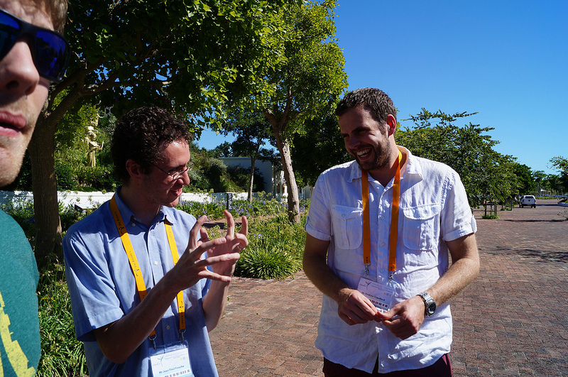
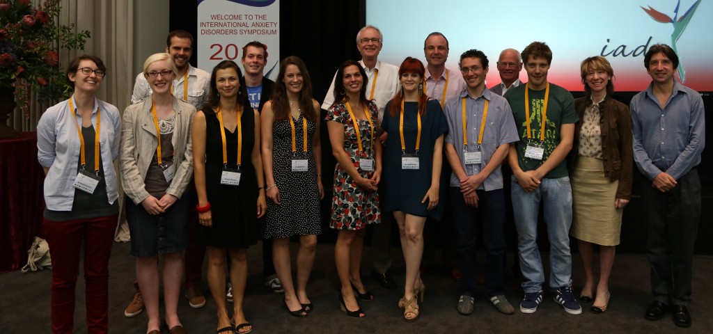
IADS 2013 was by far one of the best conferences I’ve had the pleasure of attending/presenting at, ever. Having only one speaker at any one time meant that presentations were unilaterally interesting, smartly run, well-attended and all of the speakers were right at the forefront of their respective research fields (granted, there isn’t yet a massive skin-picking field… but it’s on its way…). Tea and coffee (or a variety of refreshing produce from the free smoothie bar) was taken in the stunning backdrop of the Spier Wine Estate, with the striking Stellenbosch mountain range looming in the backdrop. Evenings were spent enjoying the local produce!But for me, the best experience of IADS was, by far, being thrust into the vibrant, surprisingly large EUSARNAD research group that had descended onto Spier. On Saturday evening, at the first EUSARNAD colloquium, established leaders in their research fields were heavily outnumbered by researchers relating to each other on the skills they’d learnt (in institutions all over the world), the relationships they’d formed and the plans they had for continuing their work. As the ‘newbie’, I found it both inspiring and challenging – this lot were clearly no slackers!
Some of the researchers were coming to the end of their respective exchange postings, and about to move on to the next challenge – so the evening was spent enjoying the scenery while they passed on tips and advice they’d learnt while here. For me, it really summed up the driving force behind the initiative: I’ve only been here for 4 days, but I’ve formed bonds with several researchers with totally different skillsets to me who are working towards the same goal – it’s really exciting for the future!
– Ben


Learning the MRI craft (one month to go) – by Ben Ainsworth

I’m off to Cape Town in a little under a month, and recently I realised I haven’t actually seen an fMRI scanner in action since my toddler-sister was unwillingly forced into one by our parents to examine her earache. She’s now 23, so I thought it might be a good idea to brush up on what actually goes on during an fMRI examination.

fMRI is one of the most exciting techniques that psychologists can use to look inside the brain. MRI stands for magnetic resonance imaging, which uses magnets to align specific atoms inside the brain in certain directions. The manner in which these atoms line up with the magnetic fields is different for each type of brain matter – MRI scanners can detect this variation and use it to produce very pretty pictures! Functional MRI scanning (fMRI) involves matching these magnetic alignments to specific brain activities – and from there, you can see which parts of the brain are active (and therefore useful) for specific thought processes. It’s all very clever.

I started by reading a few pretty helpful guides online, amongst copious amounts of technobabble (and some highly regarded articles). Because I grew up in the web-generation (we can’t actually read more than one or two lines of text before we start thinking about lunch or X-factor) I ended up watching some helpful youtube videos, as well as a couple of great TED talks.

Luckily for me, fellow Southampton psychologist Kate Sully hasn’t quite finished her experiment looking at whether brothers and sisters of ADHD sufferers have similar brain functioning patterns, and she invited me to spend a Sunday afternoon with her examining 4 teenagers down at the Southampton General Hospital.
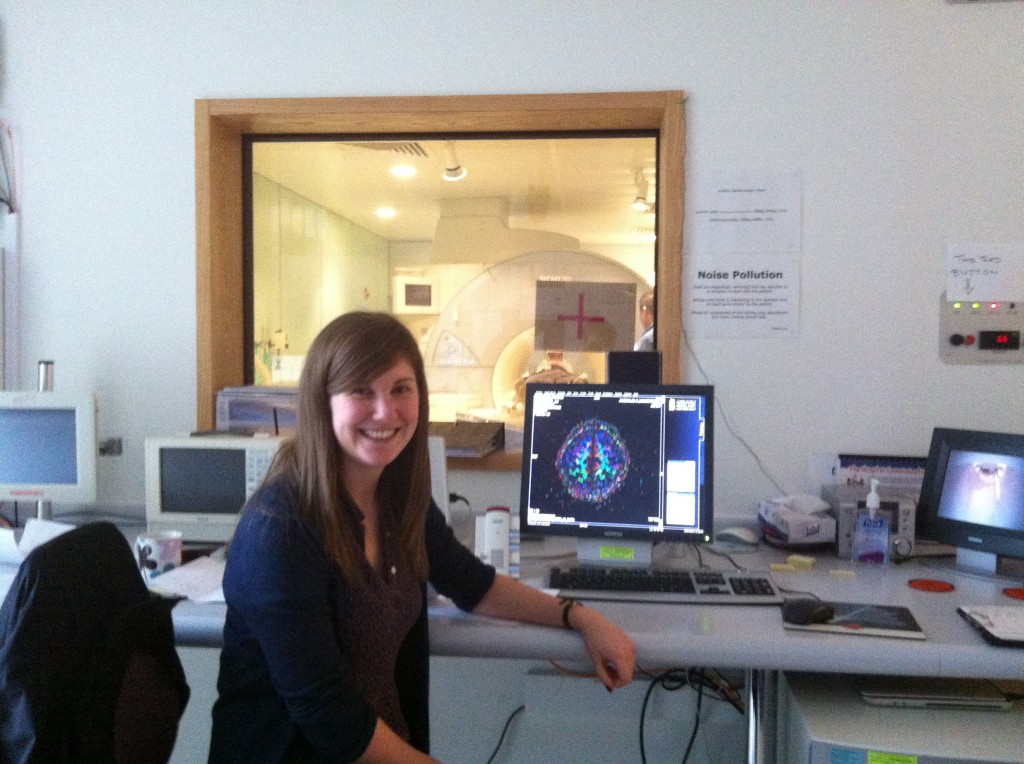
I was immediately grateful that I didn’t have to actually learn how to use the MRI machine – a very helpful radiologist (see below – the photo was taken through the doorway as MRI scanners and electronic equipment doesn’t mix too well) took some time out of his day to explain to me how everything works, and Kate took me through a lot of the protocols that are needed to ensure safe working with such a powerful (and expensive) machine.
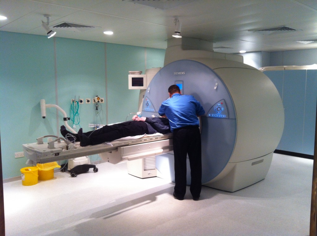
Although using the scanner seems fun, Kate has assured me that many psychologists’ roles are based around maintaining the appropriate experimental rigour. Still, it was great to see what actually goes on at the lab, particularly because I now have a much better idea of what is achievable in terms of experimental design (read: ways to squeeze an anxiety-inducing inhalation into a scanning room). Later on this week, when Kate receives CDs full of MRI pictures, I’ll be getting my teeth into some analysis.