I’m off to Cape Town in a little under a month, and recently I realised I haven’t actually seen an fMRI scanner in action since my toddler-sister was unwillingly forced into one by our parents to examine her earache. She’s now 23, so I thought it might be a good idea to brush up on what actually goes on during an fMRI examination.
fMRI is one of the most exciting techniques that psychologists can use to look inside the brain. MRI stands for magnetic resonance imaging, which uses magnets to align specific atoms inside the brain in certain directions. The manner in which these atoms line up with the magnetic fields is different for each type of brain matter – MRI scanners can detect this variation and use it to produce very pretty pictures! Functional MRI scanning (fMRI) involves matching these magnetic alignments to specific brain activities – and from there, you can see which parts of the brain are active (and therefore useful) for specific thought processes. It’s all very clever.
I started by reading a few pretty helpful guides online, amongst copious amounts of technobabble (and some highly regarded articles). Because I grew up in the web-generation (we can’t actually read more than one or two lines of text before we start thinking about lunch or X-factor) I ended up watching some helpful youtube videos, as well as a couple of great TED talks.
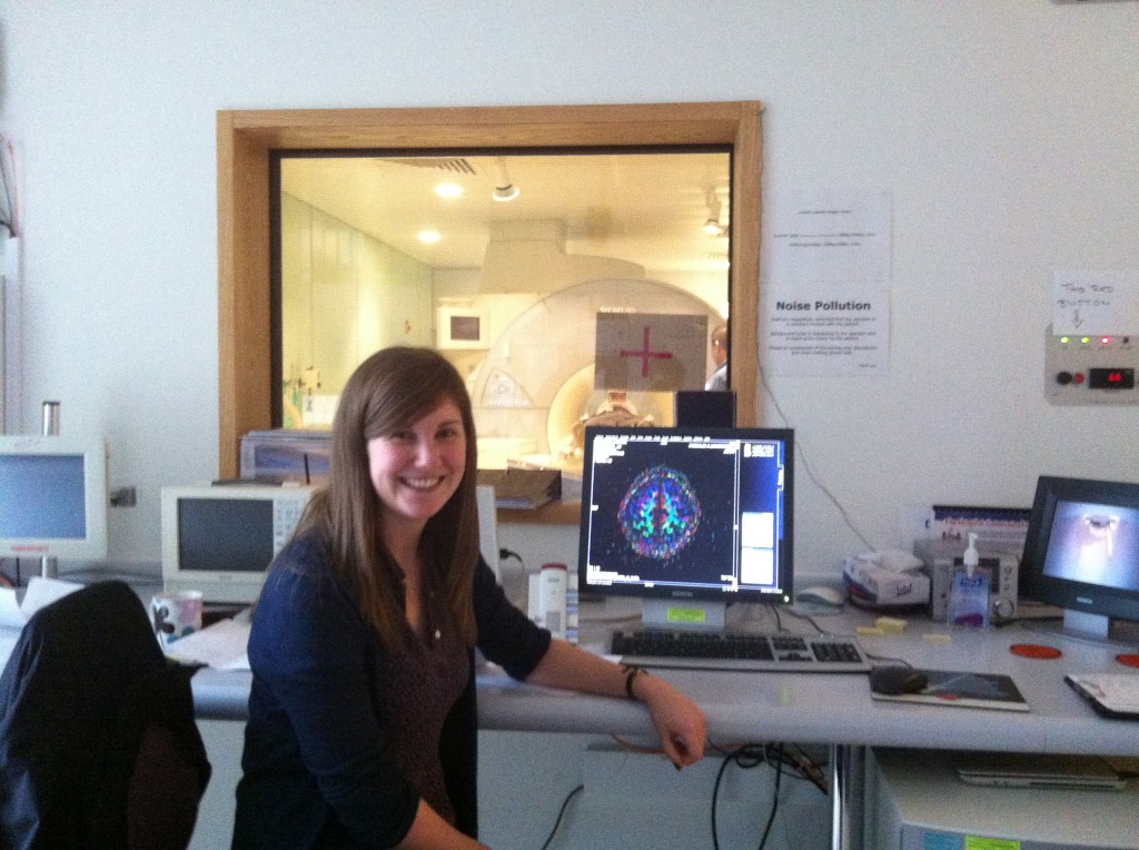
Luckily for me, fellow Southampton psychologist Kate Sully hasn’t quite finished her experiment looking at whether brothers and sisters of ADHD sufferers have similar brain functioning patterns, and she invited me to spend a Sunday afternoon with her examining 4 teenagers down at the Southampton General Hospital.
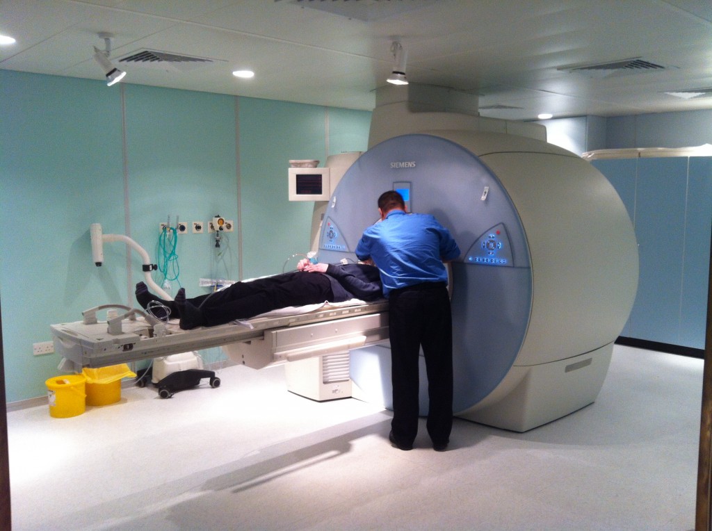
I was immediately grateful that I didn’t have to actually learn how to use the MRI machine – a very helpful radiologist (see below – the photo was taken through the doorway as MRI scanners and electronic equipment doesn’t mix too well) took some time out of his day to explain to me how everything works, and Kate took me through a lot of the protocols that are needed to ensure safe working with such a powerful (and expensive) machine.
Although using the scanner seems fun, Kate has assured me that many psychologists’ roles are based around maintaining the appropriate experimental rigour. Still, it was great to see what actually goes on at the lab, particularly because I now have a much better idea of what is achievable in terms of experimental design (read: ways to squeeze an anxiety-inducing inhalation into a scanning room). Later on this week, when Kate receives CDs full of MRI pictures, I’ll be getting my teeth into some analysis.


